The next time you sit in the chair and the hygienist takes a few X-rays, there is a good chance a piece of software looks at them before your dentist does. AI dental X-ray software is now installed in thousands of practices across the US, UK, Europe, India and Australia, drawing coloured boxes around suspected cavities and measuring bone loss down to the millimetre. It is one of the quietest AI rollouts in healthcare — and most patients have no idea it is happening.
This is not a chatbot or a gimmick. Several of these systems are cleared by the US Food and Drug Administration as medical devices, and some are used by insurers to decide whether your claim gets paid. Here is what they actually do, what the evidence says, and what it means for you.
How does AI read a dental X-ray?
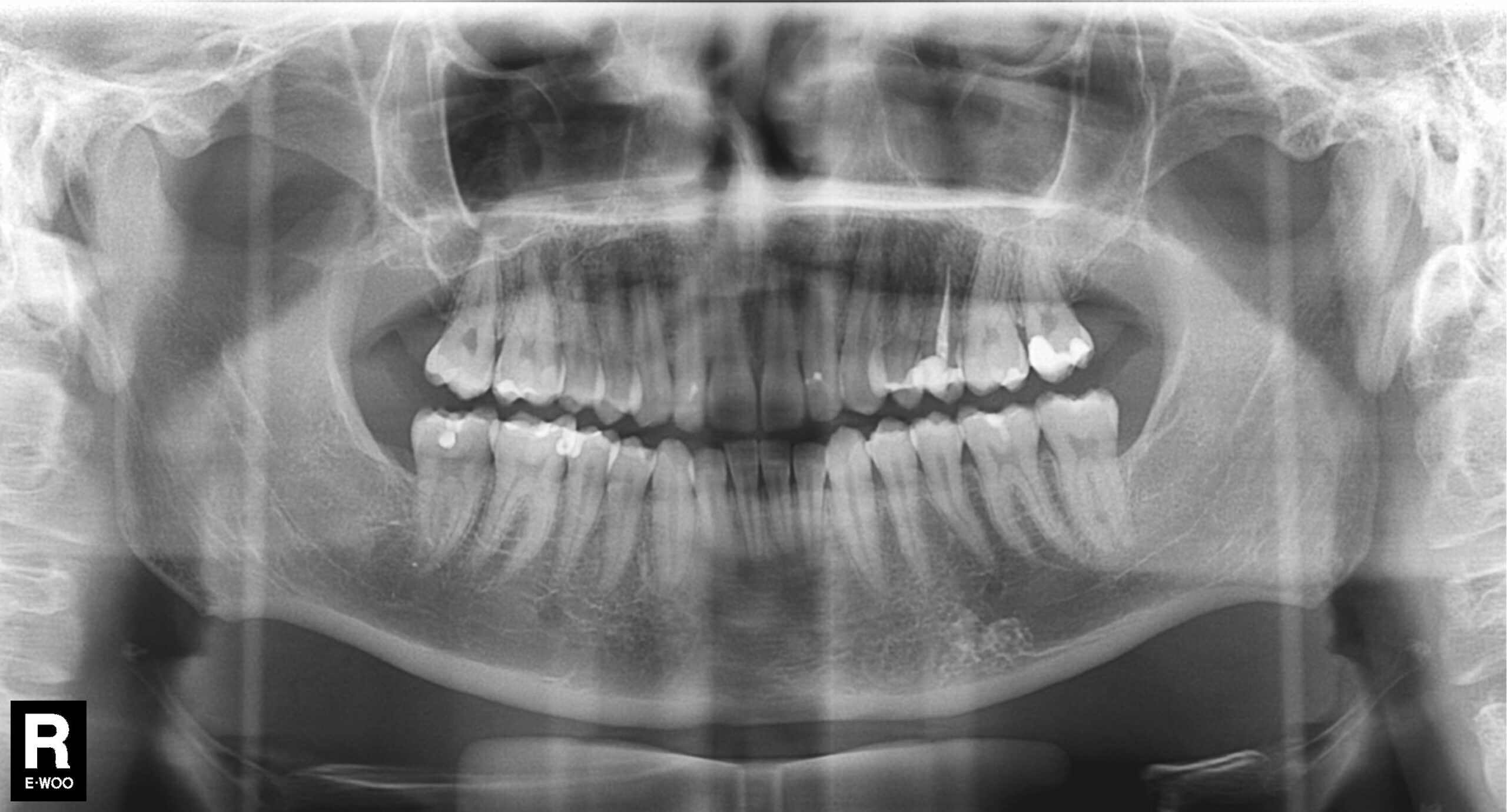
AI reads a dental X-ray using computer vision — a neural network trained on hundreds of thousands of labelled radiographs. It scans each image pixel by pixel, compares patterns against what it learned, and outlines regions that look like decay, calculus, bone loss or a failing filling. It does not diagnose; it flags findings for the dentist to confirm.
The workflow is almost invisible. The X-ray sensor sends the image to the practice management software, the AI processes it in a few seconds, and the image appears on the monitor with overlays already drawn on it. Your dentist then accepts, rejects or adjusts each finding.
In one line: dental AI is a second pair of eyes that never gets tired, never rushes the last appointment of the day, and marks up every image the same way.
What can dental AI actually detect?
The two best-known platforms are Pearl (its product is called Second Opinion) and Overjet. Both hold FDA 510(k) clearances, which is what separates them from generic image-enhancement tools. Pearl’s Second Opinion was cleared under 510(k) number K210365, and in December 2025 Pearl added clearance for detecting pathologies on panoramic radiographs. Overjet’s caries detection module was first cleared in May 2022 for bitewing X-rays and expanded in March 2023 to cover periapical radiographs.
Between them, the cleared capabilities cover:
- Caries (tooth decay) at several stages, on bitewing, periapical and panoramic images
- Calculus — hardened plaque below the gumline
- Bone loss measurement, quantified in millimetres rather than estimated by eye
- Periapical radiolucencies — dark areas at the root tip that often signal infection
- Margin discrepancies on existing crowns and fillings
- Impacted third molars (wisdom teeth) on panoramic views
- Automated charting of which teeth are present, restored or missing
Notably, Overjet holds separate clearances for caries detection in children as young as four, because a child’s developing teeth look different on film and a model trained on adults cannot be assumed to work on them.
Is AI better than a dentist at spotting cavities?
On radiographs specifically, AI models now match or beat individual dentists at detecting caries in published studies — but “better” means better at not missing things, not better at deciding what to do. A 2025 deep learning study of caries in posterior teeth reported 0.93 accuracy and 0.96 recall, meaning the model caught almost every genuine lesion while occasionally flagging healthy teeth.
That trade-off is deliberate and worth understanding. These systems are tuned toward sensitivity — they would rather raise a false alarm than let a cavity slip through. The result is that a percentage of the boxes on your X-ray will be things your dentist looks at and dismisses. That is the system working as designed, not malfunctioning.
It also explains the single biggest patient concern: does AI lead to unnecessary drilling? The honest answer is that the software creates that risk, and the safeguard against it is the clinician. A flagged early lesion may be one to monitor and remineralise rather than fill. If your dentist treats every coloured box as a treatment plan, the problem is the dentist, not the algorithm.
The same pattern shows up elsewhere in medical imaging. We covered it in detail in our piece on how AI detects diabetic retinopathy from eye scans — high sensitivity, human confirmation, and real value in catching disease early.
Why your dental insurer cares about AI X-ray reading
Here is the part patients rarely hear about. The same AI that reads X-rays in the surgery is also used on the other side of the transaction, by insurers reviewing claims.
Overjet’s platform is used by major US dental carriers including Delta Dental, and Guardian Life was an early adopter for insurance claim review. Instead of a human reviewer squinting at a submitted radiograph to judge whether the bone loss justifies the periodontal procedure billed, the AI measures it and attaches an objective number to the claim.
For patients and practices this cuts both ways. Consistent, measurable evidence means fewer arbitrary denials and faster prior authorisations. It also means a claim that does not match what the image shows is much harder to push through. The direction of travel is the same one we described in AI in emergency ambulance dispatch: the technology quietly becomes the arbiter of a decision that used to be a judgment call.
What rules govern AI in dentistry?
The American Dental Association published the first US national standard for AI in dentistry in 2025. ANSI/ADA Standard No. 1110-1:2025 sets criteria for how data from 2D radiographs should be annotated and collected so images can be exchanged and used reliably for diagnosis, treatment and research.
Alongside it, ADA Technical Report No. 1109:2025 argues that AI algorithms analysing 2D dental images should be validated against an independent dataset — one held by a third party with no ties to the AI manufacturer, based on known diagnoses. That is a pointed recommendation: it means a vendor’s own accuracy claims should not be the last word.
Running through all of it is a single principle worth remembering: AI in dentistry is a clinical decision support tool, not a replacement for clinical judgment. In the US, FDA clearance for these products is granted on exactly that basis — as an aid to the dentist reading the image.
Outside the US the picture varies: European practices work under the EU Medical Device Regulation, the UK relies on MHRA device registration, and India and Australia have growing private-clinic adoption with lighter dental-specific rules. “FDA-cleared” on a brochure tells you the product passed a bar somewhere — not that your local regulator reviewed it.
What this means when you are in the chair
You do not need to become an expert in convolutional neural networks to make good use of this. A few practical points:
- Ask to see the screen. The overlays are designed to be readable by non-specialists, and seeing your own bone loss measured is more persuasive than being told about it.
- Ask what the AI flagged versus what your dentist confirmed. These are two different lists, and the gap between them tells you how the practice uses the tool.
- Ask about watchful waiting. For an early enamel lesion, “monitor it at the next visit” is often a legitimate option.
- A flag is not a diagnosis. The software’s job is to make sure nothing gets overlooked, not to decide what happens next.
The most useful thing about AI dental X-ray software may be the least dramatic one. Radiograph reading has always varied — between dentists, between practices, and between the first appointment of the morning and the last of a long day. A system that marks up every image to the same standard removes some of that variation. That is a modest claim, and it is also the real reason this technology has spread so quickly.
Frequently Asked Questions
Is AI reading my dental X-rays without my consent?
In most jurisdictions the AI is treated as part of the imaging software your dentist uses, so there is no separate consent step. You are entitled to ask whether your practice uses it and which product. Practices that have invested in it are usually happy to show you.
Can AI replace my dentist?
No. Both the FDA clearances and the ADA’s 2025 standards frame these systems as clinical decision support — they flag findings on an image, and a licensed clinician confirms them and decides on treatment. AI cannot examine you, take a history or perform the work.
Does AI dental software cause unnecessary treatment?
It can if it is used badly. The models are deliberately tuned to catch as many real lesions as possible, which means some healthy teeth get flagged too. A good dentist dismisses those; the safeguard is professional judgment, not the software.
Which dental AI products are FDA-cleared?
Pearl’s Second Opinion (510(k) K210365) and Overjet’s detection modules are the two most widely deployed FDA-cleared platforms in the US, covering caries, calculus, bone loss and related findings across bitewing, periapical and panoramic radiographs.
Do dental insurers use AI on my X-rays too?
Yes, some do. Carriers including Delta Dental and Guardian Life use AI to review radiographs submitted with claims, measuring findings such as bone loss to assess whether the billed procedure is supported by the image.
Does AI work on children’s dental X-rays?
Only with models validated for children. Overjet holds separate FDA clearances for paediatric caries detection covering patients as young as four, because developing teeth appear differently on radiographs than adult teeth.
Sources: American Dental Association — Artificial Intelligence in Dentistry standards, Forbes coverage of AI in dentistry (July 2025).